Consider the eye to be a
camera. The camera has a system of lenses in front
which focus light on a film or digital sensors on
the back. In the same way the eye has a system of
lenses in front which focus light onto a layer at
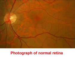
the back called the Retina. This layer has living
sensors to sense the light form and send a signal to
the brain which interprets these signals as the
image we see. The retina is connected to the brain
though a living cable which is called the optic
nerve.
Retina has a very complex structure having ten
layers. The light detector cells (photoreceptors) of
the retina are called rods and cones. Cones help
more with daylight vision and color vision whereas
rods are primarily responsible for night vision or
vision in low light conditions.
The central
part of the retina called the macula has the maximum
light sensitivity and resolution.
So any disease process which
distorts the retina or makes it thicker / thinner or
reduces the functional ability of the cells of the
retina gravely affect the vision.
To diagnose various retinal diseases certain tests
are done such as Fluorescein Angiography and Optical
Coherence Tomography. These tests and some common
retinal diseases are described in various sections.
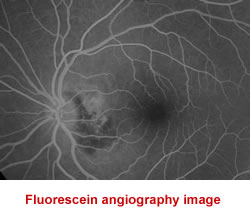
Fundus Fluorescein
Angiography ( FFA )
Fluorescein Angiography is
an investigation to further investigate the cause of
the retinal disease. In this 3 ml of a water soluble
fluorescent dye is injected into a vein on the
patient's arm. As the dye reaches the blood vessels
of the retina (takes only 10 seconds !! ),
sequential photographs are taken using a
sophisticated digital camera. The abnormal leakage
of dye or absence of normal pattern of dye gives the
doctor clues regarding the diagnosis and severity of
the retinal disease.
This test is simple with generally no significant
side-effects.
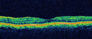
Optical Coherence
Tomography (OCT )
OCT is
a tool by which highly magnified photographs
of the retina can be taken to study it's
microscopic structure. The patient has to
just sit in front of a machine for a few
minutes and look at a target light while
these special images are acquired. It also
helps to measure the retinal thickness in
microns. So the doctor can determine if the
retina is getting thicker or thinner. Also
which layers of the retina are getting more
affected can be evaluated and the response
to treatment can be judged by serial
examinations.