| ACCOMMODATION |
The ability of the eye to change its focus
from distant to near objects; process
achieved by the lens changing its shape. |
| ANTERIOR CHAMBER |
The space in front of the iris and behind
the cornea. |
AQUEOUS HUMOR, AQUEOUS FLUID
(A-kwe-us) |
Clear, watery fluid that flows between and
nourishes the lens and the cornea; secreted
by the ciliary processes. |
| ASTIGMATISM (uh-STIG-muh-tizm) |
A
condition in which the surface of the cornea
is not spherical; causes a blurred image to
be received at the retina. |
| BLIND SPOT |
(1) A small area of the retina where the
optic nerve enters the eye; occurs normally
in all eyes.
(2) Any gap in the visual field
corresponding to a area of the retina where
no visual cells are present; associated with
eye disease. |
| CENTRAL VISION |
See VISUAL ACUITY. |
| CHOROID (KOR-oyd) |
The layer filled with blood vessels that
nourishes the retina; part of the uvea. |
| CILIARY MUSCLES |
The muscles that relax the zonules to enable
the lens to change shape for focusing. |
| CILIARY PROCESSES |
The extensions or projections of the ciliary
body that secrete aqueous humor. |
| CONES, CONE CELLS |
One type of specialized light-sensitive
cells (photoreceptors) in the retina that
provide sharp central vision and color
vision. Also see RODS. |
| CONJUNCTIVA (KAHN-junk-TY-vuh) |
The thin, moist tissue (membrane) that lines
the inner surfaces of the eyelids and the
outer surface of the sclera. |
| CONTRAST SENSITIVITY |
The ability to perceive differences between
an object and its background. |
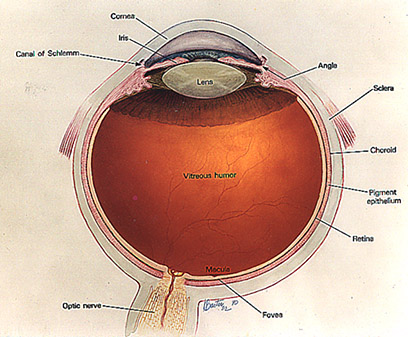
| CORNEA (KOR-nee-uh) |
The outer, transparent, dome-like structure
that covers the iris, pupil, and anterior
chamber; part of eye's focusing system. |
| DILATION |
A
process by which the pupil is temporarily
enlarged with special eye drops (mydriatic);
allows the eye care specialist to better
view the fundus. |
| FUNDUS |
The interior lining of the eyeball,
including the retina, optic disc, and
macula; portion of the inner eye that can be
seen during an eye examination by looking
through the pupil. |
| HYPEROPIA (hy-pur-OH-pee-uh) |
Farsightedness; ability to see distant
objects more clearly than close objects; may
be corrected with glasses or contact lenses. |
| INTRAOCULAR PRESSURE (IOP) |
Pressure of the fluid inside the eye; normal
IOP varies among individuals. |
| IRIS |
The colored ring of tissue suspended behind
the cornea and immediately in front of the
lens; regulates the amount of light entering
the eye by adjusting the size of the pupil. |
| LACRIMAL GLAND (LAK-rih-mul) |
The small almond-shaped structure that
produces tears; located just above the outer
corner of the eye. |
| LENS |
The transparent, double convex (outward
curve on both sides) structure suspended
between the aqueous and vitreous; helps to
focus light on the retina. |
| LEGAL BLINDNESS |
In the U.S., (1) visual acuity of 20/200 or
worse in the better eye with corrective
lenses (20/200 means that a person must be
at 20 feet from an eye chart to see what a
person with normal vision can see at 200
feet) or (2) visual field restricted to 20
degrees diameter or less (tunnel vision) in
the better eye. |
| MACULA (MAK-yoo-luh) |
The small, sensitive area of the central
retina; provides vision for fine work and
reading. |
| MYOPIA (my-OH-pee-uh) |
Nearsightedness; ability to see close
objects more clearly than distant objects;
may be corrected with glasses or contact
lenses. |
| OPTIC CUP |
The white, cup-like area in the center of
the optic disc. |
| OPTIC DISC/OPTIC NERVE HEAD |
The circular area (disc) where the optic
nerve connects to the back part of the
retina. |
| OPTIC NERVE |
The bundle of over one million nerve fibers
that carry visual messages from the retina
to the brain. |
| PERIPHERAL VISION (per-IF-ur-al) |
Side vision; ability to see objects and
movement outside of the direct line of
vision. |
| POSTERIOR CHAMBER |
The space between the back of the iris and
the front face of the vitreous; filled with
aqueous fluid. |
| PRESBYOPIA (prez-bee-OH-pee-uh) |
The gradual loss of the eye's ability to
change focus (accommodation) for seeing near
objects caused by the lens becoming less
elastic; associated with aging; occurs in
almost all people over age 45. |
| PUPIL |
The adjustable opening at the center of the
iris that allows varying amounts of light to
enter the eye. |
| RETINA (RET-in-nuh) |
The light-sensitive layer of tissue that
lines the back of the eyeball; sends visual
impulses through the optic nerve to the
brain. |
| RETINAL PIGMENT EPITHELIUM (RPE) (ep-ih-THEE-lee-um) |
The pigment cell layer that nourishes the
retinal cells; located just outside the
retina and attached to the choroid. |
| RODS, ROD CELLS |
One type of specialized light-sensitive
cells (photoreceptors) in the retina that
provide side vision and the ability to see
objects in dim light (night vision). Also
see CONES. |
| SCHLEMM'S CANAL |
The passageway for the aqueous fluid to
leave the eye. |
| SCLERA (SKLEH-ruh) |
The tough, white, outer layer (coat) of the
eyeball; with the cornea, it protects the
entire eyeball. |
| TRABECULAR MESHWORK (truh-BEC-yoo-lur) |
The spongy, mesh-like tissue near the front
of the eye that allows the aqueous fluid
(humor) to flow to Schlemm's canal then out
of the eye through ocular veins. |
| UVEA, UVEAL TRACT (YOO-vee-uh) |
The middle coat of the eyeball, consisting
of the choroid in the back of the eye and
the ciliary body and iris in the front of
the eye. |
| VISUAL ACUITY |
The ability to distinguish details and
shapes of objects; also called central
vision. |
| VISUAL FIELD |
The entire area that can be seen when the
eye is forward, including peripheral vision. |
| VITREOUS (VIT-ree-us) |
The transparent, colorless mass of gel that
lies behind lens and in front of retina. |
| ZONULES (ZAHN-yoolz) |
The fibers that hold the lens suspended in
position and enable it to change shape
during accommodation. |